
Radiology in Niš: MRI, CT scans, ultrasound and X-rays
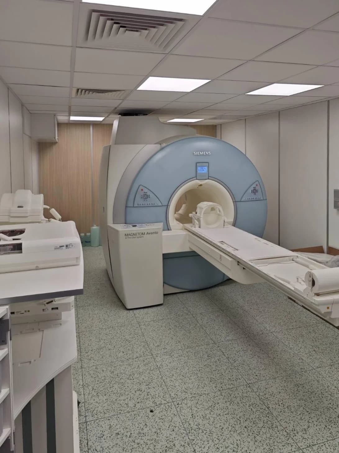
Maja Radiology in Niš, in the Maja Hospital building at Vizantijski bulevar 39, performs magnetic resonance imaging (MRI) and MSCT (CT) scans, including MSCT angiography, as well as ultrasound, colour Doppler, X-rays and bone densitometry (DEXA).
At a glance
Full description
Ultrasound scans are performed by a doctor who specialises in radiology, and images from the other methods are interpreted by a radiologist, who also writes the report. Examinations with contrast require special preparation. Prices are listed in the radiology price list, and appointments are booked by phone on 018 530 530.
Radiology: procedures
Full price list →MRI of the head and neck
Imaging of the brain, pituitary gland, eye sockets (orbits), sinuses and soft tissues of the neck. Also included is imaging of the blood vessels of the head and neck (MR angiography, with the option of venography) and of the brachial plexus, the network of nerves supplying the shoulder and arm. MRI shows soft tissues in detail, without X-ray radiation.
Read more: MRI of the head and neck →MRI of the spine, joints, arms and legs
Imaging of the cervical (neck), thoracic (chest) and lumbosacral (lower back and sacrum) spine, two spinal segments or the whole spine, as well as the hips, knee, shoulder, elbow, wrist, ankle, hand, foot, thigh and lower leg. It shows the spinal cord, discs, nerves, ligaments, tendons, cartilage, muscles and bones.
Read more: MRI of the spine, joints, arms and legs →MRI of the abdomen, pelvis, breasts and heart
Imaging of the abdominal organs, bile ducts (MRCP), small bowel (enterography), urinary tract (urography), pelvis, prostate, breasts and heart, as well as whole-body MRI. Some examinations, such as breast MRI, cardiac MRI and enterography, are most often done with contrast or special preparation. The doctor decides whether contrast is needed.
Read more: MRI of the abdomen, pelvis, breasts and heart →MSCT (CT) scans of the head, body, spine and joints
Imaging of the head, neck, chest and lungs, abdomen and pelvis, sinuses, orbits and temporal bones, kidneys and urinary tract, spine and joints. Also included are cancer staging (assessing how far a tumour has spread) and scanning of several body regions after an injury (PAN MSCT), when a doctor requests them. In the case of a serious injury, do not wait for a booked appointment: call an ambulance on 194.
Read more: MSCT (CT) scans of the head, body, spine and joints →MSCT angiography (blood vessel imaging)
Imaging of the blood vessels with contrast given into a vein: the blood vessels of the head and neck, the pulmonary arteries, the aorta and abdominal blood vessels, and the arteries of the arms and legs. It shows narrowing, widening and other changes in the blood vessels.
Read more: MSCT angiography (blood vessel imaging) →Contrast, preparation and sedation
Separate items that go with MRI and CT examinations: the contrast agent, preparation for an examination with contrast, and deep sedation during an MRI scan when the doctor considers it necessary. Whether contrast is needed is determined by the reason for the scan.
Ultrasound scans (sonography)
Examination using sound waves, without radiation: abdomen, thyroid gland, breasts, urinary and reproductive organs, prostate, soft tissues of the neck and armpits, shoulder, knee and other joints. Also included are scans most often done in babies and children: cranial ultrasound through the fontanelle (the soft spot on a baby's head), hip ultrasound and thymus ultrasound.
Read more: Ultrasound scans (sonography) →Colour Doppler scans of blood vessels and organs
An ultrasound examination of blood flow: the arteries and veins of the neck, arms and legs, as well as the blood supply to the thyroid gland, abdominal and urinary organs, pelvic organs, testicles, breasts and soft tissues. The examination involves no radiation.
Read more: Colour Doppler scans of blood vessels and organs →X-rays and bone densitometry (DEXA)
X-rays of the lungs and heart, skull, nasal bones and sinuses, spine, shoulder, hip, knee, hand, wrist and ankle, foot, individual bones, abdomen and urinary tract, as well as bone age assessment in children. Bone densitometry (DEXA) measures bone density, most often of the hip and spine, with a very low dose of radiation.
Read more: X-rays and bone densitometry (DEXA) →Our specialists: Radiology
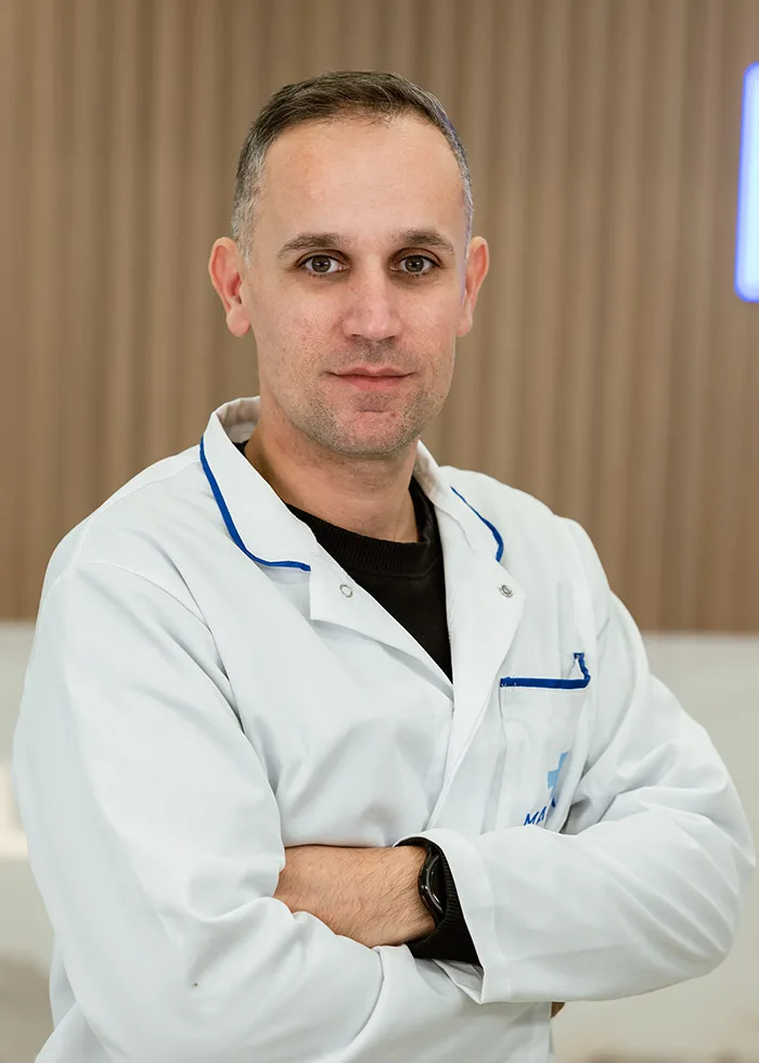

Prim. Dr Biserka Stanojević
specialist in radiology
Book an appointment with Prim. Dr Biserka Stanojević

Your visit step by step
Step 1: Your doctor's recommendation
Imaging usually starts with a recommendation or referral from the doctor treating you, stating the area to be scanned and the reason for the scan.
Step 2: Booking an appointment
Book your appointment by phone on 018 530 530.
Step 3: Preparing for the scan
Preparation depends on the examination.
Step 4: On the day of the scan
Arrive a few minutes early and check in at reception.
Step 5: Report and next steps
An ultrasound scan is performed, and its report written, by a doctor who specialises in radiology.
Show more: Your visit step by step
Your doctor's recommendation
Imaging usually starts with a recommendation or referral from the doctor treating you, stating the area to be scanned and the reason for the scan. If the same area has been scanned before, bring the report and images for comparison.
Booking an appointment
Book your appointment by phone on 018 530 530. Say which examination you need and whether your doctor has stated that contrast is needed. Tell us about a pacemaker or other implanted device, metal parts in your body, pregnancy, allergies and kidney disease. If you find it difficult to stay in an enclosed space, mention that too. If the scan is for a child, give the child's age.
Preparing for the scan
Preparation depends on the examination. For an abdominal ultrasound you usually need to fast beforehand, and for an ultrasound of the urinary organs and pelvis you need to come with a full bladder. For most MRI scans without contrast you do not need to fast, except for scans of the abdomen and bile ducts. For an examination with contrast, a recent creatinine test result is usually needed. You will be given exact instructions for your examination when you book.
On the day of the scan
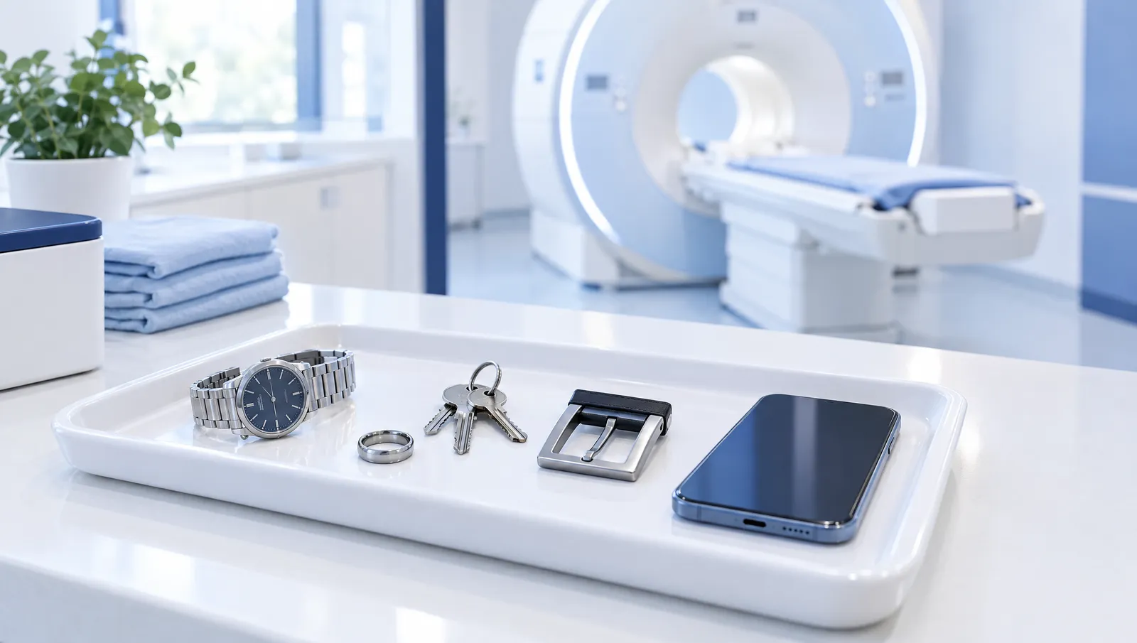
Arrive a few minutes early and check in at reception. Before an MRI scan, you will need to put away metal objects, jewellery, your watch, hairpins, hearing aid, removable dentures, bank cards and phone. Tell the staff if you have a medicated skin patch or a tattoo in the area being scanned. The MRI scanner makes loud noises while it is working, which is normal. During the scan it is important to lie still, and the procedure is explained to you before it begins.
Report and next steps
An ultrasound scan is performed, and its report written, by a doctor who specialises in radiology. After the scan, a radiologist reviews the MRI, CT and X-ray images and writes the report. Take the report to the doctor treating you or the doctor who referred you. That doctor recommends the next steps in your treatment, based on the report and an examination.
How to prepare
- Bring an identity document and a list of the medicines you take regularly.
- For an MRI scan, bring your implant card or documentation and tell us about any metal fragments, especially in the eye.
- Tell us about any previous reaction to contrast, asthma, an overactive thyroid and diabetes.
- Tell us if you are due to have treatment or a scan of the thyroid with radioactive iodine.
Show 2 more: How to prepare
- Wear comfortable clothing without metal parts.
- If your child is to be sedated, follow the food and drink instructions exactly and come with someone to accompany you.
In detail
What is radiology and what is imaging used for?
Radiology is the branch of medicine that uses images of the inside of the body to help the doctor make a diagnosis, monitor the course of treatment and plan the next steps, for example an operation. Maja Radiology in Niš performs MRI, MSCT (CT) scans, ultrasound and colour Doppler scans, X-rays and bone densitometry. Ultrasound scans are performed by a doctor who specialises in radiology, and images from the other methods are interpreted by a radiologist, who also writes the report. The report is one part of the overall assessment. The final conclusion and treatment plan are decided by the doctor treating you, after a consultation and examination.
How do MRI, CT, ultrasound and X-rays differ?
Magnetic resonance imaging (MRI) uses a strong magnetic field and radio waves, without X-ray radiation. It is most often chosen for the brain, spine, joints and soft tissues. An MSCT (CT) scanner uses X-rays and produces cross-sectional images of the body in a short time, so it is often used for the lungs, bones, abdomen and blood vessels. Ultrasound uses high-frequency sound waves, without radiation, and is most often used for the thyroid gland, abdominal organs, breasts, joints and soft tissues. A colour Doppler scan is an ultrasound examination that shows blood flow through the blood vessels. An X-ray is a quick image, most often of the bones, lungs and abdomen, and bone densitometry (DEXA) measures bone density. The doctor assesses which method is suitable, depending on the reason for the scan.
When does a doctor recommend a scan?
A scan is most often recommended by your GP or a specialist after an examination, when an abnormality needs to be checked or ruled out. Imaging is also recommended to monitor a known condition, before and after an operation, after an injury, and as part of preventive check-ups when a doctor recommends them. The reason for the scan determines which area is scanned and whether contrast is needed. That is why it is useful to bring your referral or your doctor's recommendation and any previous reports.
Radiology at the same address as Maja Hospital and Maja Health Centre
Maja Radiology is at Vizantijski bulevar 39 in Niš, in the building that houses Maja Hospital and Maja Health Centre. The laboratory and the pharmacy are at the same address. When your doctor asks for blood tests as well as a scan, for example before an examination with contrast, the tests can be done in the same building. Take the radiologist's report to the doctor treating you or the doctor who referred you, whether they work within the Maja Medical group or at another healthcare facility.
Frequently asked questions
Do I need a referral for an MRI or CT scan?
X-rays and CT scans use X-ray radiation. Under radiation protection rules, a doctor assesses whether such a scan is justified. That is why you should bring a referral or recommendation from your doctor, stating the area to be scanned and the reason for the scan. For MRI and ultrasound, a doctor's recommendation is also useful, because the radiologist uses it to choose the scanning technique and to assess whether contrast is needed. Check when booking whether you can come for your examination without a referral.
Which is better, an MRI or a CT scan?
No single method is right for every examination. The choice depends on the reason for the scan and the doctor's assessment. MRI uses a magnetic field and radio waves, without X-ray radiation, and shows soft tissues in detail, such as the brain, spinal cord, discs, ligaments and cartilage. A CT scan (MSCT) uses X-rays, takes less time and is often chosen for the lungs, bones, injuries and blood vessels. In the case of sudden and severe symptoms, do not wait for a booked appointment: call an ambulance on 194.
How much radiation is there in a CT scan or an X-ray?
X-rays and MSCT (CT) scans use X-ray radiation. The dose from a plain X-ray is low, and the dose from a CT scan is higher, because a large number of cross-sectional images are taken. Bone densitometry uses a very low dose. That is why a scan is done when the doctor judges that the benefit of the result outweighs the risk, and for children a method without radiation is chosen whenever possible. Always tell us before the scan if you are or might be pregnant. Ultrasound and MRI do not use X-ray radiation.
What happens during a scan with contrast?
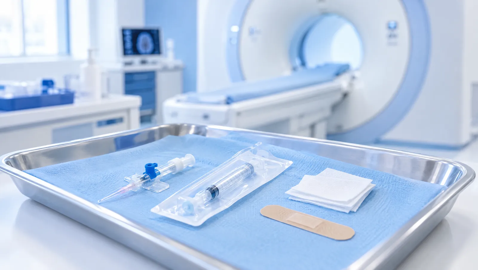
The contrast agent is most often given into a vein in the arm during the scan, to show the blood vessels and certain abnormalities more clearly. Different types of contrast are used for MRI and for CT. During a CT scan, a brief feeling of warmth in the body or a metallic taste in the mouth is common while the contrast is being given. Before the examination, a recent creatinine test, which shows how well the kidneys are working, is usually needed, and in severe kidney disease contrast is given only after a doctor's assessment. You will be told when you book whether you need to fast. Tell us in advance about allergies, previous reactions to contrast, asthma, kidney disease, an overactive thyroid and diabetes. If you notice a rash, itching or swelling after the examination, contact a doctor, and if you have difficulty breathing, call an ambulance on 194. The doctor assesses whether contrast is needed.
Are scans also done for children?
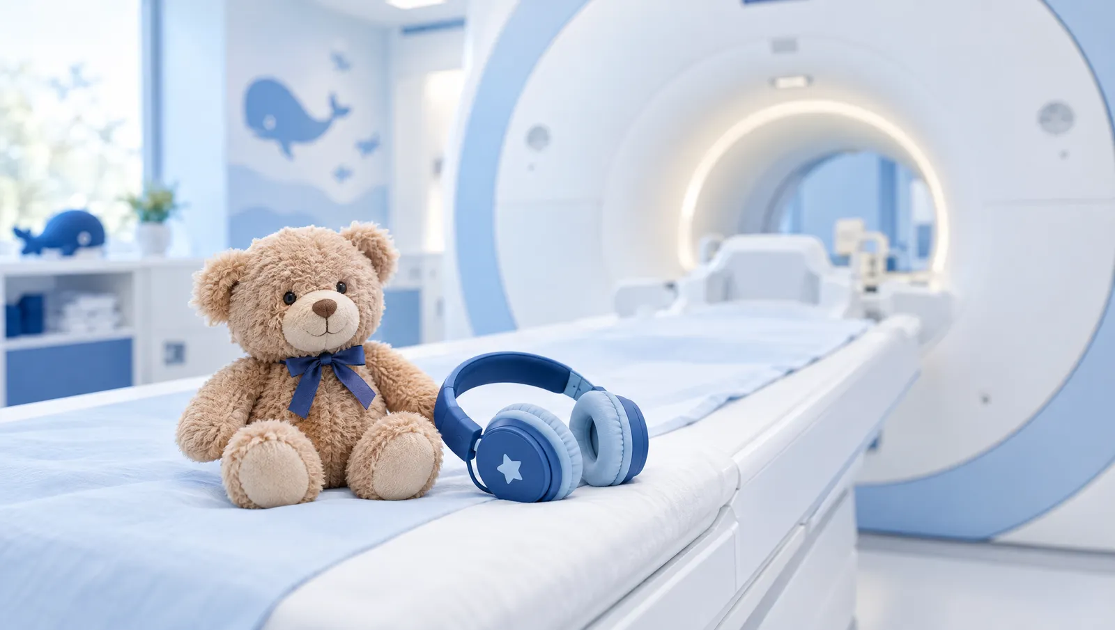
The radiology price list includes examinations most often done in babies and children, such as cranial ultrasound through the fontanelle, hip ultrasound and X-ray bone age assessment. For children, ultrasound and MRI are preferred whenever possible, because they do not use X-ray radiation. For an MRI scan, the patient has to lie still for quite a long time, which is often difficult for young children. Check when booking whether your child's examination can be done and how. Give your child's age and the reason for the scan, so that the examination can be properly planned.
Show 2 more: Frequently asked questions
How much does a scan cost at Maja Radiology?
The price depends on the type of examination, the area being scanned and whether contrast is given. In the price list, contrast, preparation for an examination with contrast and deep sedation during an MRI scan are listed as separate items. Check when booking whether any of these will be added to the price of your examination. Prices of examinations, from an X-ray to a whole-body MRI, are in the radiology price list and next to each group of examinations on this page.
How do I book a scan and when will the report be ready?
Scans are booked by phone on 018 530 530. When booking, say which area is to be scanned, whether contrast is needed and whether you have implanted metal parts or devices. The time needed for the report depends on the type of examination. Ask when booking or on the day of the scan when and how you can collect your report.
Advice and answers
Procedures
Colour Doppler
Read more →
Procedures
MRI scan (magnetic resonance imaging)
Read more →
Procedures
MSCT scan (CT scan)
Read more →
Procedures
X-ray and bone densitometry
Read more →
Procedures
Ultrasound
Read more →
Advice
MRI and claustrophobia: how to prepare for your scan
Advice
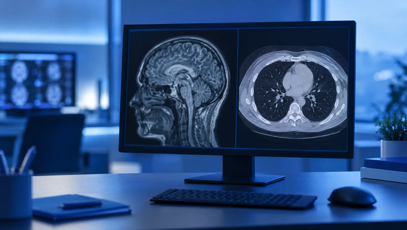
MRI or CT scan: what is the difference and who decides which scan you need
Advice
MRI for children: preparation, sedation and having a parent present
Advice
MRI with contrast: when it is needed, preparation and blood tests before the scan
Advice
Preparing for an MRI scan: what to wear, what to bring and how long the scan takes
The information in this text is general in nature and does not replace an examination by a doctor.
Book an appointment: Radiology
Information on this website does not replace a medical examination. Treatment is decided by a doctor after the examination.