MRI or CT scan: what is the difference and who decides which scan you need
An MRI scan and a CT scan (MSCT, CT) both produce detailed cross-sectional images of the body, but in different ways. MRI uses a magnetic field and radio waves, with no X-ray radiation; it takes longer and shows soft tissues in more detail. A CT scan uses X-rays, the scan itself takes a few minutes, and it is most often chosen for bones, the lungs and injuries. Neither scan is better at everything: the doctor who refers you decides which one you need. Both scans are carried out at Maja Radiology, in the Maja Hospital building in Niš.
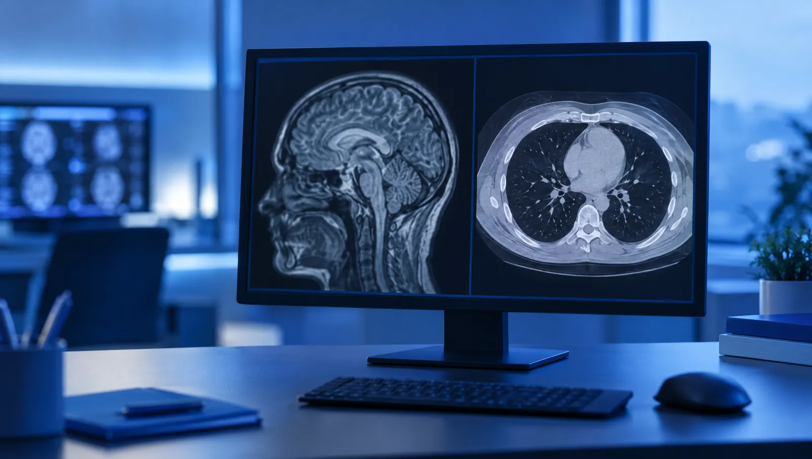
How does an MRI scan work, and how does a CT scan work?
Both scans produce cross-sectional images of the body, similar to thin slices. A specialist radiologist reviews these images on a computer and then writes the report. The difference lies in how the images are obtained.
Magnetic resonance imaging (MR, MRI) uses a strong magnetic field and radio waves. Different tissues in the body give off different signals, and a computer builds an image from these signals. MRI does not use X-rays, that is, ionising radiation.
In reports and price lists, this scan is called CT or MSCT. CT stands for computed tomography, and MSCT for multislice computed tomography, in which the machine captures several slices in a single rotation. You may also hear the term multislice scanner. A CT scanner uses X-rays: an X-ray tube rotates around the body, and a computer uses the data to create cross-sectional images and, where needed, three-dimensional images of bones and blood vessels.
How long does the scan take and what is it like inside the machine?
Patients notice the biggest difference in how long the scan takes and in the machine itself:
- Duration: an MRI scan of one area usually takes 20 to 45 minutes. The CT scan itself usually takes a few minutes. With both scans, you will be there longer if contrast is given.
- Space: with most MRI machines, you lie on a table that slides into a longer tunnel, with the part of the body being scanned in the middle of the tunnel. A CT scanner has a short, wide ring that the table moves slowly through, so it usually feels less enclosed.
- Noise: an MRI machine makes loud knocking and buzzing sounds while it is working, so patients are usually given earplugs or headphones. A CT scanner is much quieter.
- Keeping still: with both scans it is important to keep still, but for longer with MRI. When the chest and abdomen are scanned, you may sometimes be asked by the radiographer to hold your breath briefly, both for CT and for MRI.
Because of the length of the scan and the tunnel of the machine, MRI is more demanding for young children, for people who find it hard to lie still for a long time and for those who feel uncomfortable in enclosed spaces. If this applies to you, tell the doctor who refers you and mention it when you book.
What does an MRI scan show better, and what does a CT scan show better?
The question "which is better" does not have a single answer. Each scan has areas where it provides more information. This is a general division, and in practice the choice depends on the question the doctor is asking and on previous results. The abdomen, for example, is scanned with both methods, depending on the organ and on what the doctor wants to check.
MRI is most often chosen for soft tissues:
- the brain, the pituitary gland and the spinal cord
- the discs and nerve roots in the spine
- ligaments, tendons, menisci and cartilage in the joints
- the pelvic organs, the prostate, the liver and the bile ducts
- the breasts and the heart, when the doctor requests such a scan
- the blood vessels of the head and neck (MR angiography)
A CT scan is most often chosen when speed matters, or for imaging bones or the lungs:
- the lungs and the organs of the chest
- fractures and complex injuries of bones and joints
- assessment after a head injury, when it is important to see bleeding or a fracture quickly
- blood vessels, with contrast injected into a vein (CT angiography), for example the aorta and the blood vessels of the lungs, arms and legs
- the sinuses, and stones in the kidneys and urinary tract
- scanning several areas of the body at once, for example when assessing how far a disease has spread
CT or MRI scan of the head?
The head is a common example of this question. A CT scan of the head is quick and shows the bones of the skull and recent bleeding well. That is why, in emergencies, for example after a fall or a blow to the head, a CT scan is often done first.
Emergencies are handled by the emergency ambulance service, not by a booked appointment. If, after a blow to the head, there is loss of consciousness, vomiting, a severe headache, confusion or drowsiness, or if there is sudden weakness in an arm or leg, difficulty speaking or a drooping face, do not book an appointment; call an ambulance on 194 without delay.
An MRI scan of the head shows brain tissue in more detail. Doctors most often choose it for smaller changes in the brain, for examining the pituitary gland, for monitoring changes over time and when a CT scan does not provide enough information. Some head scans also need contrast, which the radiologist decides on in line with the doctor's referral.
A similar principle applies to the spine. MRI shows the discs, nerves and spinal cord more clearly, while a CT scan shows the bony parts of the vertebrae and fractures.
Radiation, metal in the body and pregnancy: what should you look out for?
MRI does not use ionising radiation. A magnetic field and radio waves are not the same type of radiation as X-rays. That is why doctors often choose MRI for repeated scans and for children, when it provides the information needed.
A CT scan uses X-rays. The dose depends on the area and the scanning protocol, and it is higher than for an ordinary X-ray. The scan is done when the doctor judges that the benefit of the result outweighs the possible risk. So bring your previous scans with you, so that the scan is not repeated unnecessarily.
With MRI, the main safety question is metal in the body. Before the scan, you must tell staff if you have a pacemaker, an implanted defibrillator, a cochlear implant, a neurostimulator, an insulin pump or another implanted device, as well as metal clips on blood vessels, a metal fragment in the eye, shrapnel or any other metal in the body. With many implants, MRI is possible under certain conditions, and the radiologist decides on the basis of the implant's documentation. For pacemakers and defibrillators, a cardiologist is usually also involved in the assessment.
For a CT scan, metal in the body is usually not an obstacle, but it can make the image less clear in that part of the body. So before a CT scan, also tell staff which implants or devices you have.
If a scan with contrast is planned, for both MRI and CT, tell staff if you have had a reaction to contrast before or if you have kidney disease. Tell staff if you are pregnant or might be pregnant, both before an MRI scan and before a CT scan.
Who decides which scan you need?
The scan is chosen by the doctor who refers you: your GP or a specialist, for example a neurologist, orthopaedic surgeon, surgeon or urologist. They know what needs to be checked and what previous results show. That is why the referral or recommendation should state the area to be scanned and the reason for the scan.
The radiologist then uses the referral to choose the scanning protocol and to assess whether contrast is needed. If another method is better suited to the doctor's question, the radiologist may suggest it, in agreement with the referring doctor. Sometimes both scans are needed, one after the other, because they provide different information.
That is why it is not a good idea to choose a scan only because it seems more precise or because you can get an appointment sooner. A scan that does not answer the doctor's question may mean another scan and additional cost. If it is not clear to you why that particular scan has been recommended, ask the doctor who is referring you.
How do you book an MRI or CT scan at Maja Radiology?
According to the radiology price list, both MRI and MSCT scans are carried out at Maja Radiology, in the Maja Hospital building at Vizantijski bulevar 39 in Niš: head, spine, joints, abdomen, pelvis and blood vessels. MRI is also used to scan the breasts and the heart, and CT to scan the chest and lungs. Appointments are booked by phone on 018 530 530.
When booking, give the exact name of the scan as stated on the doctor's referral or recommendation. Bring with you to the scan:
- the doctor's referral or recommendation
- previous reports and scans of the same area
- your implant card or hospital discharge summary, if you have an implanted device
- a creatinine test result, if contrast is planned and if the instructions you receive when booking ask for it
The price depends on the type of scan and the area being scanned. The price list has separate items for contrast and for preparation for a scan with contrast. Check what exactly is included in the price of your scan when you book. Prices for MRI and CT scans of the head, as well as the items for contrast, are shown in the table below, and prices for all scans can be found in the radiology price list.
The information in this text is general in nature and does not replace an examination by a doctor.